
The basic idea here is to recolonize your gut with a large amount of specific probiotics and nutrients. This work presents a new approach of biofilm sensing in the early attachment stage with a low limit of detection up to 104 RIU refractive index units or 35 20 103 CFUmL colony formed units.

Biofilm Formation Assay Kit Dojindo
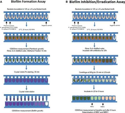
The early biofilm concentration was determined by crystal violet CV binding assay.
. In place of inverting and discarding the media which can lead to the loss of the aerobic biofilm layer in CV assay media was removed from the formed biofilm with the help of a syringe and biofilm layer was allowed to dry. Microtitre plate assay for biofilm quantification. However a number of other colorimetric and metabolic stains have been reported for the quantification of biofilm formation using the.
Your goal for this step is to Restore the gut microbiome using friendly bacteria and the building blocks your body requires in order to stabilize towards long-term health. In summary the protocol optimized by ImQuest BioSciences provides a high throughput procedure to screen drug candidates for activity against P. MBEC assay is a very time efficient and accurate.
Take out 3-4 100ul aliquots of EtOH. Step 3 is the last and my favorite step. In the protocol described here we focus on the use of 96-well optically clear polystyrene flat-bottom plate to study biofilm formation by Leptospira spp.
In this assay the extent of biofilm formation is measured using the dye crystal violet CV. Inoculate 5ml liquid medium with 5µl 1st Overnight culture use disposable test tubes and incubate at proper conditions overnight. In this assay 5 mm glass- or.
3 The peg lid is placed in the bacteria culture and incubated to generate the biofilm. Apply the second overnight culture into plates as your experimental designed pattern and perform the experiment as follow. The seven Rs Relevance lab outcome field outcome Reasonableness expense lab techniques Resemblance controls similar between exps.
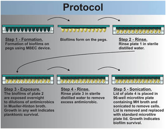
The MBEC Assay Biofilm Inoculators consist of a plastic lid with 96 pegs and one of two corresponding bases. Current standardized efficacy testing protocols of disinfectants however employ predominantly planktonic bacteria. Timing 3 d i Establish biofilms in 96-well plates.
It is a Protocol Extension of our previous Nature Protocols article which describes the synthesis of SPOT-peptide arrays and assays for. In the proposed protocol loss of such biofilm layer was prevented. 4 The peg lid is gently rinsed to removed planktonic bacteria and a serial diluted test solution is dispensed into a new 96-well microplate.
In this assay the extent of biofilm formation is measured using the dye crystal violet CV. The MBEC assay uses a Calgary Biofilm Device CBD a 96-well plate with pegs built into the lid that allow for the adherence and growth of biofilm. V329 biofilm was formed on pre-sterilized 96 well flat bottom.
We also describe an alternative method based on phase contrast image analysis that we believe is more suitable for accurately quantifying. 4- keep without no agitation for 24 or 48 or 72 days until. Let biofilms air dry 45min room temp 3.
Briefly A 10 µl of cell. The staining and washing steps were avoided and an organic solvent-tetrahydrofuran. Make sure that the only crystal.
Remove media from biofilms and wash 1X in 1ml PBS 2. Stain all wells used in the assay with 125 μL of 01 crystal violet for 10 minutes. Aeruginosa biofilms and is especially suitable to rapidly identify highly active inhibitors of biofilm formation.
Suspension having 05 OD. Because it utilizes a 96-well plate format it is suitable as a tool for screening large numbers of bacterial strains or species. One base contains 96 individual wells Figure 1.
Shake the 96-well plate over the tray again and rinse out the crystal violet in a large beaker of water. The MBEC Minimum Biofilm Eradication Concentration Assay is a high throughput screening assay used to determine the efficacy of antimicrobials against biofilms of a variety of microorganisms. The microtiter plate biofilm assay see Basic Protocol 1 is a useful method for assessing bacterial attachment by measuring the staining of the adherent biomass.
Initial attachment followed by irreversible attachment maturation and dispersion. Attributes of a standard method. Remove Crystal Violet stain 5.
Polystyrene micro-titre plates in triplicates as described elsewhere. In order to test the efficacy of biocides on biofilms in a standardized manner a new assay was developed and optimized for easy-handling quickness low running costs and above allrepeatability. In the protocol described here we will focus on the use of this assay to study biofilm formation by the model organism Pseudomonas aeruginosa.
Add 1ml 04 Crystal Violet stain to each biofilm and let sit room temp 45min 4. 5 The pegs covered in biofilm are. Was inoculated in 190 µl TSB medium in each well and 200 µl.
Kwasny SM and Opperman TJ. A step-by-step summary of the MBEC Assay. 1- grow the bacteria 2- guarantee the pure culture 3- Next form the biofime add 500 microLiters of bacteria into a 24 well microplate.
The MBEC minimum biofilm eradication concentration Assay can be used to determine the efficacy of an antimicrobial agent against biofilm. Static biofilm cultures of gram-positive pathogens grown in a microtiter for-. Wash 4X with 3ml H2O gently to remove unbound stain 6.
1-2 Bacteria culture is prepared and dispensed into a 96-well microplate. The matrix is made by the microorganisms within the biofilm throughout. However a number of other colorimetric and metabolic stains have been reported for the quantification of biofilm formation using the.
In the protocol described here we will focus on the use of this assay to study biofilm formation by the model organism Pseudomonas aeruginosa. Biofilm Assay Protocol for Biofilm assay by Safranin using 96-well plates. Cover the bench top with more paper towels and hit the plate against the bench top until all wells are free of liquid crystal violet.
Add 2ml 100 EtOH to each biofilm and let sit 45 min room temp 7. The air-liquid interface ALI assay see Basic Protocol 2 is complementary. And quantify the biofilm formation by crystal violet CV staining.
Biofilm eradication assay. There are 4 stages of biofilm formation. Label two identical 96-well microtiter plates following one of the outlines supplied in.
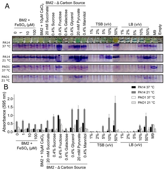
However such a concentration was lower than the detection limit of this assay.
Antimicrobial Sensitivity

Biofilm Formation Assay In Pseudomonas Syringae Bio Protocol

Assess The Cell Viability Of Staphylococcus Aureus Biofilms Thermo Fisher Scientific Ca

Quantification Of Biofilm Biomass By Staining Non Toxic Safranin Can Replace The Popular Crystal Violet Sciencedirect

A Combination Of Phenotype Microarraytm Technology With The Atp Assay Determines The Nutritional Dependence Of Escherichia Coli Biofilm Biomass Intechopen

Optimized Protocol For H Volcanii Immersed Liquid Biofilm Formation Download Scientific Diagram

Biofilm Formation Assay Kit Dojindo

Schematic Representation Of The Steps Involved In The Protocol For Download Scientific Diagram
Frontiers Biofilm And Planktonic Antibiotic Resistance In Patients With Acute Exacerbation Of Chronic Rhinosinusitis Cellular And Infection Microbiology

A Cartoon Of The Methodology Presented In This Protocol Biofi Lm Download Scientific Diagram
Biomolecules Free Full Text Critical Assessment Of Methods To Quantify Biofilm Growth And Evaluate Antibiofilm Activity Of Host Defence Peptides Html

An Overview Of The High Throughput Protocol For Metal Susceptibility Download Scientific Diagram

Biofilm Eradication Testing For Antimicrobial Efficacy

Polystyrene Microtiter Plate Biofilm Assay Of A Pleuropneumoniae Download Scientific Diagram

Microtiter Dish Biofilm Formation Assay Protocol

Biofilm Formation Assay Kit Testpiece Dojindo Eu
2
2

Biofilm Formation Assay Kit Dojindo